CD279 (PD-1) Monoclonal Antibody (J43), Brilliant Ultra Violet™ 805, eBioscience™, Invitrogen™
Manufacturer: Fischer Scientific
Select a Size
| Pack Size | SKU | Availability | Price |
|---|---|---|---|
| Each of 1 | 36-899-8582-Each-of-1 | In Stock | ₹ 36,579.00 |
36-899-8582 - Each of 1
In Stock
Quantity
1
Base Price: ₹ 36,579.00
GST (18%): ₹ 6,584.22
Total Price: ₹ 43,163.22
Antigen
CD279 (PD-1)
Classification
Monoclonal
Concentration
0.2 mg/mL
Formulation
PBS with BSA and 0.09% sodium azide; pH 7.2
Gene Accession No.
Q02242
Gene Symbols
Pdcd1
Purification Method
Affinity chromatography
Regulatory Status
RUO
Gene ID (Entrez)
18566
Content And Storage
4° C, store in dark, DO NOT FREEZE!
Form
Liquid
Applications
Flow Cytometry
Clone
J43
Conjugate
Brilliant Ultraviolet 805
Gene
Pdcd1
Gene Alias
CD279; EGK_05005; hPD1; hPD-1; hPD-l; hSLE1; Ly101; mPD-1; PD1; PD-1; Pdc1; Pdcd1; programmed cell death 1; programmed cell death 1 protein; programmed cell death protein 1; programmed cell death protein 1-like; programmed death 1; Protein PD1; protein PD-1; sCD279; SLEB2; soluble CD279; systemic lupus erythematosus susceptibility 2
Host Species
Armenian Hamster
Quantity
100 μg
Primary or Secondary
Primary
Target Species
Mouse
Product Type
Antibody
Isotype
IgG
Related Products

Description
- The J43 monoclonal antibody reacts with mouse PD-1 (programmed death-1), a 55 kDa member of the Ig superfamily
- PD-1 contains the ITIM and plays a key role in peripheral tolerance and autoimmune disease in mice
- PD-1 is expressed mainly on activated T and B lymphocytes
- Two novel B7 Family members have been identified as PD-1 ligands, PD-L1 (B7-H1) and PD-L2 (B7-DC)
- Evidence reported to date suggests overlapping functions for these ligands and their constitutive expression on some normal tissues and upregulation on activated antigen-presenting cells
- It is reported that J43 inhibits the binding of mouse PD-L1-Ig and mouse PD-L2-Ig to PD-1/BHK transfected cells
- When administrated in vivo, both intact and Fab of J43 are reported to enhance contact hypersensitivity and exacerbate acute GVHD similar to transfer of PD-1-deficient cells
- Injection of J43 also exacerbates EAE and NOD diabetes as do specific antibodies to mouse PD-L1 and PD-L2
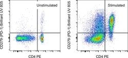
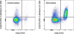
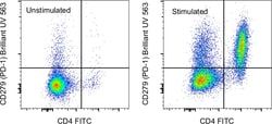
- Applications Reported: This J43 antibody has been reported for use in flow cytometric analysis
- Applications Tested: This J43 antibody has been tested by flow cytometric analysis of stimulated mouse splenocytes
- This may be used at less than or equal to 0.5 μg per test
- Cell number should be determined empirically but can range from 10^5 to 10^8 cells/test
- Brilliant Ultraviolet 805 is a tandem dye that emits at 797 nm and is intended for use on cytometers equipped with an ultraviolet (355 nm) laser
- Cell-mediated immune responses are initiated by T lymphocytes that are themselves stimulated by cognate peptides bound to MHC molecules on antig en-presenting cells (APC)
- T-cell activation is generally self-limited as activated T cells express receptors such as PD-1 (also known as PDCD-1) that mediate inhibitory signals from the APC
- PD-1 can bind two different but related ligands, PDL-1 and PDL-2
- Upon binding to either of these ligands, signals generated by PD-1 inhibit the activation of the immune response in the absence of "edanger signals"e such as LPS or other molecules associated with bacteria or other pathogens
- Evidence for this is seen in PD1-null mice who exhibit hyperactivated immune systems and autoimmune diseases
- Despite its predicted molecular weight, PD-1 often migrates at higher molecular weight in SDS-PAGE.