CD49b (Integrin alpha 2) Monoclonal Antibody (DX5), PE-Cyanine7, eBioscience™, Invitrogen™
Manufacturer: Fischer Scientific
Select a Size
| Pack Size | SKU | Availability | Price |
|---|---|---|---|
| Each | 50-155-99-Each | In Stock | ₹ 25,511.85 |
50-155-99 - Each
In Stock
Quantity
1
Base Price: ₹ 25,511.85
GST (18%): ₹ 4,592.133
Total Price: ₹ 30,103.983
Antigen
CD49b (Integrin alpha 2)
Classification
Monoclonal
Concentration
0.2 mg/mL
Formulation
PBS with 0.09% sodium azide; pH 7.2
Gene Accession No.
Q62469
Gene Symbols
Itga2
Purification Method
Affinity chromatography
Regulatory Status
RUO
Gene ID (Entrez)
16398
Content And Storage
4° C, store in dark, DO NOT FREEZE!
Form
Liquid
Applications
Flow Cytometry
Clone
DX5
Conjugate
PE-Cyanine7
Gene
Itga2
Gene Alias
alpha 2 subunit of VLA-2 receptor; BDPLT9; BR; CD49 antigen-like family member B; CD49B; Collagen receptor; DX5; GPIa; HPA-5; human platelet alloantigen system 5; integrin alpha 2; integrin alpha-2; integrin subunit alpha 2; integrin, alpha 2; integrin, alpha 2 (CD49B, alpha 2 subunit of VLA-2 receptor); Itga2; platelet antigen Br; platelet glycoprotein GPIa; platelet membrane glycoprotein Ia; very late activation protein 2 receptor, alpha-2 subunit; VLA-2; VLA2 receptor; VLA-2 receptor, alpha 2 subunit; VLA-2 subunit alpha; VLAA2
Host Species
Rat
Quantity
100 μg
Primary or Secondary
Primary
Target Species
Mouse
Product Type
Antibody
Isotype
IgM κ
Related Products


Description
- Description: The DX5 monoclonal antibody reacts with CD49b, an antigen expressed on a majority of mouse natural killer cells and a subset of T cells
- DX5 reacts with all strains of mouse tested including the most commonly used strains, BALB/c, C57BL/6, C3H, CBA, DBA, AKR, SJL and 129
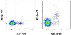
- Simultaneous staining of C57BL/6 spleen cells with anti-NK1.1 mAb (PK136) and DX5 reveals coexpression of both markers by a majority of cells as well as presence of small populations of DX5+PK136- and DX5-PK136+ cells
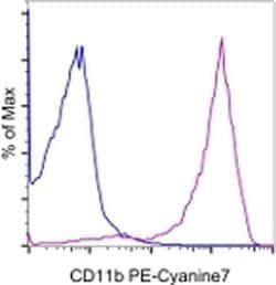
- Applications Reported: This DX5 antibody has been reported for use in flow cytometric analysis
- Applications Tested: This DX5 antibody has been tested by flow cytometric analysis of mouse splenocytes
- This can be used at less than or equal to 0.5 μg per test
- A test is defined as the amount (μg) of antibody that will stain a cell sample in a final volume of 100 μL
- Cell number should be determined empirically but can range from 10^5 to 10^8 cells/test
- It is recommended that the antibody be carefully titrated for optimal performance in the assay of interest
- Light sensitivity: This tandem dye is sensitive photo-induced oxidation
- Please protect this vial and stained samples from light
- Fixation: Samples can be stored in IC Fixation Buffer (cat
- 00-8222) (100 μL cell sample + 100 μL IC Fixation Buffer) or 1-step Fix/Lyse Solution (cat
- 00-5333) for up to 3 days in the dark at 4°C with minimal impact on brightness and FRET efficiency/compensation
- CD49b (Integrin alpha 2, Integrin alpha 2 chain, VLA-2 alpha chain, HM alpha 2) is a member of the integrin family
- It is a glycoprotein with molecular weight of 150 kD, and it complexes with CD29 (Integrin beta 1) to form the heterodimeric integrin VLA-2 (integrin alpha 2 beta 1, or GPIa/IIa) complex
- VLA-2 is an extracellular receptor for laminin, collagen, and fibronectin, and interaction with its ligands results in the activation of intracellular signaling pathways
- It has reported roles in VEGF-induced angiogenesis in vivo, as well as adhesion and lymphocyte activation
- CD49b is expressed by NK cells, NK-T cells, monocytes, platelets, and epithelial cells
- It is also expressed on adaptive immune cells such as T and B cells, specifically on a subset of CD4+ T cells in the spleen, on intraepithelial and lamina propria lymphocytes in the intestine, as well as on a population of peripheral CD4+ type 1 T regulatory (Tr1) cells that co-express LAG-3.