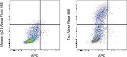
Tau Monoclonal Antibody (HT7), Alexa Fluor™ 647, eBioscience™, Invitrogen™
Manufacturer: Invitrogen
Select a Size
| Pack Size | SKU | Availability | Price |
|---|---|---|---|
| Each of 1 | 51-591-642-Each-of-1 | In Stock | ₹ 40,940.00 |
51-591-642 - Each of 1
In Stock
Quantity
1
Base Price: ₹ 40,940.00
GST (18%): ₹ 7,369.20
Total Price: ₹ 48,309.20
Antigen
Tau
Classification
Monoclonal
Concentration
5 μL/Test
Formulation
PBS with 0.2% BSA and 0.09% sodium azide, pH 7.2
Gene Accession No.
P10636
Gene Symbols
MAPT
Immunogen
Purified Human Tau protein
Quantity
100 Tests
Primary or Secondary
Primary
Target Species
Human
Product Type
Antibody
Isotype
IgG1 κ
Applications
Flow Cytometry
Clone
HT7
Conjugate
Alexa Fluor 647
Gene
MAPT
Gene Alias
AI413597, AW045860, DDPAC, FLJ31424, FTDP17, FTDP-17, G protein beta1/gamma2 subunit-interacting factor 1, map tau, Mapt, MAPTL, MGC138549, microtubule associated protein tau, microtubule-associated protein tau, microtubule-associated protein tau, isoform 4, microtubules, MSTD, Mtapt, MTBT1, MTBT2, Neurofibrillary tangle protein, neurofibrillary tangles, Neuronal Marker, paired helical filament-tau, PHFtau, PHF-tau, PPND, PPP1R103, protein phosphatase 1, regulatory subunit 103, pTau, RNPTAU, Tau, Tau microtubule-associated protein, tau protein, Tau-4, Tau5, Unknown (protein for MGC:134287)
Host Species
Mouse
Purification Method
Affinity chromatography
Regulatory Status
RUO
Gene ID (Entrez)
4137
Content And Storage
4° C, store in dark, DO NOT FREEZE!
Form
Liquid
Related Products

Description
- Description: This HT7 monoclonal antibody recognizes human and bovine Tau protein
- The epitope of this antibody has been mapped to residues 159 through 163, corresponding to the amino acid sequence PPGQK of human Tau40
- In certain cell lines and tissue other than brain, clone HT7 may show a low but distinct amount of nonspecific staining
- Therefore we recommend the use of clone HT7 to detect Tau expression by flow cytometry in human Tau transfected cells or tissue derived from human Tau transgenic mice
- This antibody does not cross-react with murine Tau
- Applications Reported: This HT7 antibody has been reported for use in flow cytometric analysis
- Applications Tested: This HT7 antibody has been pre-diluted and tested by flow cytometric analysis of Human Tau transfected 293 HEK cells using the Intracellular Fixation & Permeabilization Buffer Set (Product No
- 88-8824-00) and protocol
- Please refer to Staining Intracellular Antigens for Flow Cytometry, Protocol A: Two step protocol for intracellular (cytoplasmic) proteins located at www.thermofisher.com/flowprotocols
- This antibody may be used at 5μL (0.25 μg) per test
- A test is defined as the amount (μg) of antibody that will stain a cell sample in a final volume of 100 μL
- Cell number should be determined empirically but can range from 10^5 to 10^8 cells/test
- Tau is a neuronal microtubule-associated protein found predominantly on axons
- The function of Tau is to promote tubulin polymerization and stabilize microtubules
- The C-terminus binds axonal microtubules while the N- terminus binds neural plasma membrane components, suggesting that tau functions as a linker protein between both
- Axonal polarity is predetermined by TAU/MAPT localization (in the neuronal cell) in the domain of the cell body defined by the centrosome
- The short isoforms allow plasticity of the cytoskeleton while the longer isoforms may preferentially play a role in its stabilization
- In its hyper-phosphorylated form, Tau is the major component of paired helical filaments (PHF), the building block of neurofibrillary lesions in Alzheimer's diseases (AD) brain
- Hyper-phosphorylation impairs the microtubule binding function of Tau, resulting in the destabilization of microtubules in AD brains, μgtimately leading to the degeneration of the affected neurons
- Numerous serine/threonine kinases phosphorylate Tau, including GSK-3beta, protein kinase A (PKA), cyclin-dependent kinase 5 (cdk5) and casein kinase II
- Hyper-phosphorylated Tau is found in neurofibrillary lesions in a range and other central nervous system disorders such as Pick's disease, frontotemporal dementia, cortico-basal degeneration and progressive supranuclear palsy.