Ki-67 Monoclonal Antibody (SolA15), Alexa Fluor™ 532, eBioscience™, Invitrogen™
Manufacturer: Invitrogen
Select a Size
| Pack Size | SKU | Availability | Price |
|---|---|---|---|
| Each of 1 | 58-569-882-Each-of-1 | In Stock | ₹ 17,088.00 |
58-569-882 - Each of 1
In Stock
Quantity
1
Base Price: ₹ 17,088.00
GST (18%): ₹ 3,075.84
Total Price: ₹ 20,163.84
Antigen
Ki-67
Classification
Monoclonal
Concentration
0.2 mg/mL
Formulation
PBS with 0.09% sodium azide, pH 7.2
Gene Accession No.
E9PVX6, P46013
Gene Symbols
Mki67
Purification Method
Affinity chromatography
Regulatory Status
RUO
Gene ID (Entrez)
100686578, 102135895, 17345, 291234, 4288
Content And Storage
4° C, store in dark, DO NOT FREEZE!
Form
Liquid
Applications
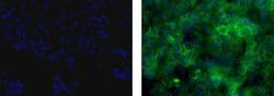
Flow Cytometry, Immunocytochemistry, Immunohistochemistry (Paraffin)
Clone
SolA15
Conjugate
Alexa Fluor 532
Gene
Mki67
Gene Alias
antigen identified by monoclonal antibody Ki 67, antigen identified by monoclonal antibody Ki-67, Antigen identified by monoclonal antibody Ki-67 homolog, Antigen KI-67, Antigen KI-67 homolog, antigen KI-67, proliferation marker protein Ki-67, antigen KI-67-like, cb31, D630048A14Rik, I79_022666, Ki67, Ki-67, KIA, LOW QUALITY PROTEIN: proliferation marker protein Ki-67, marker of proliferation Ki-67, MIB-, MIB-1, Mki67, PPP1R105, Proliferation marker protein Ki-67, proliferation-related Ki-67 antigen, protein phosphatase 1, regulatory subunit 105, RP11-380J17.2, sb:cb31, si:ch211-250b22.7, unnamed protein product, wu:fa11g09, wu:fb57a07, wu:fi14e05
Host Species
Rat
Quantity
100 μg
Primary or Secondary
Primary
Target Species
Canine, Cynomolgus Monkey, Human, Mouse, Non-human Primate, Rat
Product Type
Antibody
Isotype
IgG2a κ
Related Products


Description
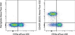
- Description: The monoclonal antibody SolA15 recognizes mouse and rat Ki-67, a 300 kDa nuclear protein
- Ki-67 is present during all active phases of the cell cycle (G1, S, G2, and mitosis), but is absent from resting cells (G0)
- Ki-67 is detected within the nucleus during interphase but redistributes to the chromosomes during mitosis
- Ki-67 is used as a marker for determining the growth fraction of a given population of cells
- In studies of tumor cells, the Ki-67 labeling index refers to the number of Ki-67 positive cells within the population and this is used to predict outcome of particular cancer types
- Ki-67 has been shown to interact with the DNA-bound protein chromobox protein homolog 3 (CBX3) (heterochromatin)
- The SolA15 antibody also recognizes human, non-human primate and canine Ki-67
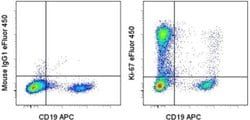
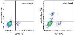
- Applications Reported: This SOLA15 antibody has been reported for use in flow cytometric analysis
- Applications Tested: This SOLA15 antibody has been tested by flow cytometric analysis of stimulated mouse splenocytes
- This may be used at less than or equal to 0.25 μg per test
- A test is defined as the amount (μg) of antibody that will stain a cell sample in a final volume of 100 μL
- Cell number should be determined empirically but can range from 10^5 to 10^8 cells/test
- It is recommended that the antibody be carefully titrated for optimal performance in the assay of interest
- Alexa Fluor 532 is excited with the Green laser (532 nm) and emits at 561 nm
- Ki-67 is a nuclear protein that is expressed during various stages in the cell cycle, particularly during late G1, S, G2, and M phases
- The protein has a forkhead associated domain (FHA) through which it associates with euchromatin at the perichromosomal layer, the centromeric heterochromatin, and the nucleolus
- Ki-67 is shown to have a cell cycle dependent topographical distribution with perinucleolar expression at G1, expression in the nuclear matrix at G2, and expression on the chromosomes during M phase
- Ki-67 is commonly used as a proliferation marker because it is not detected in G0 cells, but increases steadily from G1 through mitosis
- Ki-67 antibodies are useful in establishing the cell growing fraction in neoplasms
- In neoplastic tissues, the prognostic value is comparable to the tritiated thymidine-labelling index
- The correlation between low Ki-67 index and histologically low-grade tumors is strong
- Ki-67 is routinely used as a neuronal marker of cell cycling and proliferation.