CD56 (NCAM) Monoclonal Antibody (TULY56), Super Bright™ 600, eBioscience™, Invitrogen™
Manufacturer: Invitrogen
Select a Size
| Pack Size | SKU | Availability | Price |
|---|---|---|---|
| Each of 1 | 63-056-642-Each-of-1 | In Stock | ₹ 33,642.00 |
63-056-642 - Each of 1
In Stock
Quantity
1
Base Price: ₹ 33,642.00
GST (18%): ₹ 6,055.56
Total Price: ₹ 39,697.56
Antigen
CD56 (NCAM)
Classification
Monoclonal
Concentration
5 μL/Test
Formulation
PBS with BSA and 0.09% sodium azide
Gene Accession No.
P13591
Gene Symbols
Ncam1
Purification Method
Affinity chromatography
Regulatory Status
RUO
Gene ID (Entrez)
4684, 693789
Content And Storage
4° C, store in dark, DO NOT FREEZE!
Form
Liquid
Applications
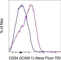
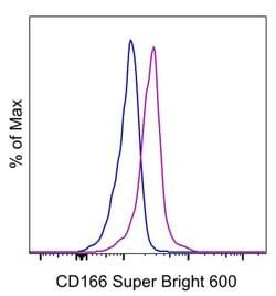
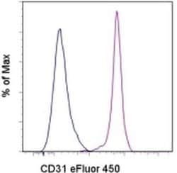
Flow Cytometry
Clone
TULY56
Conjugate
Super Bright 600
Gene
Ncam1
Gene Alias
adhesion molecule; antigen recognized by monoclonal antibody 5.1H11; Cd56; CD-56; CD56 120 kDa GPI-linked isoform; CD56 140 kDa isoform; CD56 140 kDa VASE isoform; E NCAM; E-NCAM; MSK39; N CAM1; NCAM; N-CAM; Ncam1; N-CAM-1; NCAM-1; NCAMC; NCAM-C; neural cell adhesion molecule; Neural cell adhesion molecule 1; neural cell adhesion molecule, NCAM; sCD56; sNCAM; soluble CD56; soluble NCAM
Host Species
Mouse
Quantity
100 Tests
Primary or Secondary
Primary
Target Species
Human, Non-human Primate, Rhesus Monkey
Product Type
Antibody
Isotype
IgG1 κ
Related Products


Description
- This TULY56 monoclonal antibody reacts with human CD56, also known as Neural Cell Adhesion Molecule (NCAM)
- CD56 is a highly glycosylated transmembrane molecule expressed by neurons and plays a role in the homotypic adhesion of neural cells
- In the hematopoietic system, CD56 is expressed on NK cells and a subset of T cells referred to as NKT cells
- Staining with TULY56 does not block binding of CMSSB, suggesting that the two antibodies recognize different epitopes
- Additionally, TULY56 performs better after fixation and permeabilization than CMSSB
- The TULY56 monoclonal antibody crossreacts with Rhesus macaque
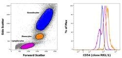
- This TULY56 antibody has been pre-titrated and tested by flow cytometric analysis of normal human peripheral blood cells
- This can be used at 5 μL (0.06 μg) per test
- A test is defined as the amount (μg) of antibody that will stain a cell sample in a final volume of 100 μL
- Cell number should be determined empirically but can range from 10^5 to 10^8 cells/test
- Super Bright 600 is a tandem dye that can be excited with the violet laser line (405 nm) and emits at 600 nm
- We recommend using a 610/20 bandpass filter
- Please make sure that your instrument is capable of detecting this fluorochrome
- When using two or more Super Bright dye-conjugated antibodies in a staining panel, it is recommended to use Super Bright Complete Staining Buffer (Product No
- SB-4401) to minimize any non-specific polymer interactions
- CD56 (NCAM, neural cell adhesion molecule) is a transmembrane glycoprotein of the immunoglobulin family that serves as an adhesive molecule and is ubiquitously expressed in the nervous system in isoforms ranging from 120-180 kDa
- CD56 is found on T cells and NK cells, and is involved in cell migration, axonal growth, pathfinding and synaptic plasticity
- Polysialic modification results in reduction of CD56-mediated cell adhesion
- Through its extracellular region, CD56 mediates homophilic and heterophilic interactions by binding extracellular matrix components such as laminin and integrins
- CD56 is expressed on most neuroectodermal derived cell lines, tissues and neoplasms such as retinoblastoma, medulloblastoma, astrocytomas and neuroblastoma
- Further, CD56 is a widely used neuroendocrine marker with a high sensitivity for neuroendocrine tumors and ovarian granulosa cell tumors
- Diseases associated with CD56 dysfunction include rabies and blastic plasmacytoid dendritic cell neoplasms.