7069222
CD279 (PD-1) Monoclonal Antibody (J43), Biotin, eBioscience™, Invitrogen™
Armenian Hamster Monoclonal Antibody
Manufacturer: Fischer Scientific
The price for this product is unavailable. Please request a quote
Antigen
CD279 (PD-1)
Concentration
0.5 mg/mL
Classification
Monoclonal
Form
Liquid
Regulatory Status
RUO
Gene Accession No.
Q02242
Gene Symbols
Pdcd1
Primary or Secondary
Primary
Content And Storage
4° C, store in dark, DO NOT FREEZE!
Gene
Pdcd1
Clone
J43
Applications
Flow Cytometry
Conjugate
Biotin
Host Species
Armenian Hamster
Target Species
Mouse
Gene ID (Entrez)
18566
Isotype
IgG
Purification Method
Affinity chromatography
Product Type
Antibody
Related Products


Description
- Description: The J43 monoclonal antibody reacts with mouse PD-1 (programmed death-1), a 55 kDa member of the Ig superfamily
- PD-1 contains the immunoreceptor tyrosine-based inhibitory motif (ITIM) and plays a key role in peripheral tolerance and autoimmune disease in mice
- PD-1 is expressed mainly on activated T and B lymphocytes
- Two novel B7 Family members have been identified as PD-1 ligands, PD-L1 (B7-H1) and PD-L2 (B7-DC)
- Evidence reported to date suggests overlapping functions for these ligands and their constitutive expression on some normal tissues and upregulation on activated antigen-presenting cells
- It is reported that J43 inhibits the binding of mouse PD-L1-Ig and mouse PD-L2-Ig to PD-1/BHK transfected cells
- When administrated in vivo, both intact and Fab of J43 are reported to enhance contact hypersensitivity and exacerbate acute GVHD similar to transfer of PD-1-deficient cells
- Injection of J43 also exacerbates EAE and NOD diabetes as do specific antibodies to mouse PD-L1 and PD-L2
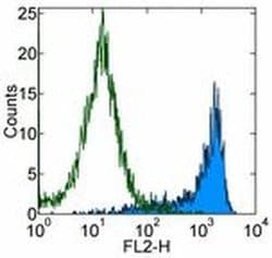
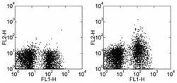
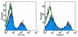
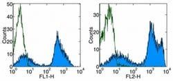
- Applications Reported: The J43 antibody has been reported for use in flow cytometric analysis
- Applications Tested: The J43 antibody has been tested by flow cytometric analysis of Con A-simulated mouse splenocytes and mouse PD-1 transfected cells
- This can be used at less than or equal to 0.25 μg per test
- A test is defined as the amount (μg) of antibody that will stain a cell sample in a final volume of 100 μL
- Cell-mediated immune responses are initiated by T lymphocytes that are themselves stimulated by cognate peptides bound to MHC molecules on antig en-presenting cells (APC)
- T-cell activation is generally self-limited as activated T cells express receptors such as PD-1 (also known as PDCD-1) that mediate inhibitory signals from the APC
- PD-1 can bind two different but related ligands, PDL-1 and PDL-2
- Upon binding to either of these ligands, signals generated by PD-1 inhibit the activation of the immune response in the absence of "edanger signals"e such as LPS or other molecules associated with bacteria or other pathogens
- Evidence for this is seen in PD1-null mice who exhibit hyperactivated immune systems and autoimmune diseases
- Despite its predicted molecular weight, PD-1 often migrates at higher molecular weight in SDS-PAGE.