Fibroblasts/Epithelial cells Antibody (D7-FIB), Allophycocyanin/Cy7, Novus Biologicals™
Manufacturer: Novus Biologicals
Select a Size
| Pack Size | SKU | Availability | Price |
|---|---|---|---|
| Each of 1 | N60777APCY7-Each-of-1 | In Stock | ₹ 66,171.50 |
N60777APCY7 - Each of 1
In Stock
Quantity
1
Base Price: ₹ 66,171.50
GST (18%): ₹ 11,910.87
Total Price: ₹ 78,082.37
Antigen
Fibroblasts/Epithelial cells
Classification
Monoclonal
Conjugate
APC-Cyanine7
Formulation
PBS with 0.05% Sodium Azide
Immunogen
Human foreskin fibroblasts
Quantity
0.1 mL
Test Specificity
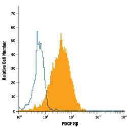
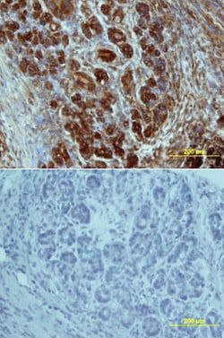
NB600-777 recognizes a 112kD molecule expressed on the cell surface of human fibroblasts and epithelial cells. In peripheral blood the antibody stains myeloid cells and a very small number of lymphocytes. Studies upon the antigen have shown it to be sensitive to SDS, but resistant to trypsin, tunicamycin and collagenase. In immunohistological studies the antibody has also been found to bind to epithelium, myoepthelium, smooth muscle and some leucocytes. D7-FIB has been shown to be useful as a cell membrane marker to characterize chondrocyte differentiation giving a positive reaction with dedifferentiated human chondrocytes, and negative with differentiated chondrocytes (3). This product is routinely tested in flow cytometry on the KG1 cell line.
Content And Storage
Store at 4°C in the dark. Do not freeze.
Applications
Flow Cytometry
Clone
D7-FIB
Dilution
Flow Cytometry
Host Species
Mouse
Purification Method
Protein G purified
Primary or Secondary
Primary
Target Species
Human
Isotype
IgG2a
Related Products


Description
- Fibroblasts/Epithelial cells Monoclonal specifically detects Fibroblasts/Epithelial cells in Human samples
- It is validated for Flow Cytometry.