V23201
Invitrogen™ Chromatin Condensation & Membrane Permeability Dead Cell Apoptosis Kit with Hoechst 33342, YO-PRO™-1, and PI dyes, for flow cytometry
Manufacturer: Invitrogen™
Select a Size
| Pack Size | SKU | Availability | Price |
|---|---|---|---|
| Each of 1 | V23201-Each-of-1 | In Stock | ₹ 34,977.00 |
V23201 - Each of 1
In Stock
Quantity
1
Base Price: ₹ 34,977.00
GST (18%): ₹ 6,295.86
Total Price: ₹ 41,272.86
Quantity
1 kit
Product Type
Dead Cell Apoptosis Kit
Flow Cytometer Laser Lines
488 nm
No. of Reactions
200 Reactions
Product Line
Vybrant™, YO-PRO™
Conjugate
Hoechst 33342, Propidium Iodide, YO-PRO-1
Form
Solution
Format
Kit
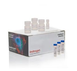
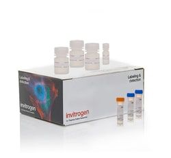
Content And Storage
Contains 1 vial of Hoechst 33342, 1 vial of YO-PRO™-1, and propidium iodide.Store in refrigerator (2–8°C) and protect from light.
Excitation/Emission
YO-PRO™-1: 491/509 cm, Hoechst 33342: 350/461 cm, PI: 535/617 cm
Shipping Condition
Room Temperature
For Use With (Application)
Flow Cytometry
For Use With (Equipment)
Flow Cytometer
Related Products


Description
- This product detects apoptotic cells with changes in nuclear chromatin condensation and plasma membrane permeability, using three nucleic acid stains: UV-excitable Hoechst 33342, green fluorescent YO-PRO™ dye, and propidium iodide
- The YO-PRO™ dye can enter apoptotic cells, whereas red-fluorescent propidium iodide (PI) cannot
- Thus after staining with YO-PRO™-1 dye and PI, apoptotic cells show green fluorescence, and dead cells show primarily red fluorescence and some green fluorescence
- Blue fluorescent Hoechst 33342 brightly stains the condensed chromatin of apoptotic cells and more dimly stains the normal chromatin of live cells
- The staining pattern resulting from the simultaneous use of these three dyes makes it possible to distinguish normal, apoptotic and dead cell populations by flow cytometry or fluorescence microscopy.