DR3 Monoclonal Antibody (JD3), eBioscience™, Invitrogen™
Manufacturer: Invitrogen
Select a Size
| Pack Size | SKU | Availability | Price |
|---|---|---|---|
| Each of 1 | 14-660-337-Each-of-1 | In Stock | ₹ 1,22,108.00 |
14-660-337 - Each of 1
In Stock
Quantity
1
Base Price: ₹ 1,22,108.00
GST (18%): ₹ 21,979.44
Total Price: ₹ 1,44,087.44
Antigen
DR3
Classification
Monoclonal
Concentration
0.5 mg/mL
Formulation
PBS with 0.09% sodium azide; pH 7.2
Gene Accession No.
Q93038
Gene Symbols
TNFRSF25
Purification Method
Affinity chromatography
Regulatory Status
RUO
Gene ID (Entrez)
8718
Content And Storage
4° C
Form
Liquid
Applications
Flow Cytometry
Clone
JD3
Conjugate
Unconjugated
Gene
TNFRSF25
Gene Alias
APO3; APO-3; apoptosis inducing receptor; apoptosis-inducing receptor AIR; Apoptosis-mediating receptor DR3; apoptosis-mediating receptor TRAMP; DDR3; death domain receptor 3 soluble form; Death receptor 3; death receptor beta; DR3; LARD; lymphocyte-associated receptor of death; Protein WSL; Protein WSL-1; RP4-650H14.2; TNF receptor superfamily member 25; TNFRSF12; TNFRSF25; TR3; TRAMP; tumor necrosis factor receptor superfamily member 25; tumor necrosis factor receptor superfamily, member 12; tumor necrosis factor receptor superfamily, member 12 (translocating chain-association membrane protein); tumor necrosis factor receptor superfamily, member 25; UNQ455/PRO779; Wsl; WSL1; WSL-1; WSL-LR
Host Species
Mouse
Quantity
2 mg
Primary or Secondary
Primary
Target Species
Human
Product Type
Antibody
Isotype
IgG1 κ
Related Products
Description
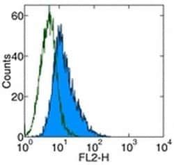
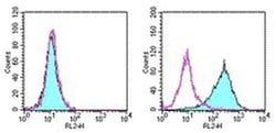
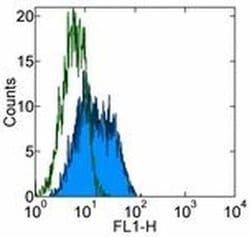
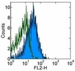
- Description: The JD3 monoclonal antibody reacts with human DR3, also known as Apo-3, WSL-1, TRAMP, LARD, DDR3, and TR3
- DR3, a novel death receptor is expressed at low level by monocytes and granulocytes
- Interaction of DR3 with its ligand activates NF-kappa B pathway and induces apoptosis
- Initially, DR3 was thought to be a receptor for TWEAK, but further studies have shown that TWEAK could induce apoptosis via receptors distinct from DR3
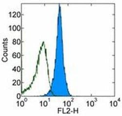
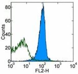
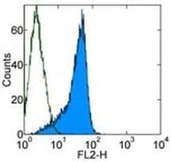
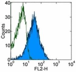
- JD3 has been reported for use in flow cytometric analysis
- Applications Reported: The JD3 antibody has been reported for use in flow cytometric analysis
- Applications Tested: The JD3 antibody has been tested by flow cytometric analysis of normal human peripheral blood cells and human DR3-transfected cells
- This can be used at less than or equal to 1 μg per test
- A test is defined as the amount (μg) of antibody that will stain a cell sample in a final volume of 100 μL
- Cell number should be determined empirically but can range from 10^5 to 10^8 cells/test
- It is recommended that the antibody be carefully titrated for optimal performance in the assay of interest
- Purity: Greater than 90%, as determined by SDS-PAGE
- Aggregation: Less than 10%, as determined by HPLC
- Filtration: 0.2 μm post-manufacturing filtered
- DR3/TNFRSF25 is a member of the TNF-receptor superfamily
- This receptor is expressed preferentially in the tissues enriched in lymphocytes, and it may play a role in regulating lymphocyte homeostasis
- This receptor has been shown to stimulate NF-kappa B activity and regulate cell apoptosis
- The signal transduction of this receptor is mediated by various death domain containing adaptor proteins
- Knockout studies in mice suggested the role of DR3/TNFRSF25 in the removal of self-reactive T cells in the thymus
- Multiple alternatively spliced transcript variants of this gene encoding distinct isoforms have been reported, most of which are potentially secreted molecules
- The alternative splicing of this gene in B and T cells encounters a programmed change upon T-cell activation, which predominantly produces full-length, membrane bound isoforms, and is thought to be involved in controlling lymphocyte proliferation induced by T-cell activation.