Ki-67 Monoclonal Antibody (SolA15), PE-Cyanine5, eBioscience™, Invitrogen™
Manufacturer: Invitrogen
Select a Size
| Pack Size | SKU | Availability | Price |
|---|---|---|---|
| Each of 1 | 15-569-882-Each-of-1 | In Stock | ₹ 37,558.00 |
15-569-882 - Each of 1
In Stock
Quantity
1
Base Price: ₹ 37,558.00
GST (18%): ₹ 6,760.44
Total Price: ₹ 44,318.44
Antigen
Ki-67
Classification
Monoclonal
Concentration
0.2 mg/mL
Formulation
PBS with 0.09% sodium azide, pH 7.2
Gene Accession No.
E9PVX6, P46013
Gene Symbols
Mki67
Purification Method
Affinity chromatography
Regulatory Status
RUO
Gene ID (Entrez)
100686578, 102135895, 17345, 291234, 4288
Content And Storage
4° C, store in dark, DO NOT FREEZE!
Form
Liquid
Applications
Flow Cytometry
Clone
SolA15
Conjugate
PE-Cyanine5
Gene
Mki67
Gene Alias
antigen identified by monoclonal antibody Ki 67, antigen identified by monoclonal antibody Ki-67, Antigen identified by monoclonal antibody Ki-67 homolog, Antigen KI-67, Antigen KI-67 homolog, antigen KI-67, proliferation marker protein Ki-67, antigen KI-67-like, cb31, D630048A14Rik, I79_022666, Ki67, Ki-67, KIA, LOW QUALITY PROTEIN: proliferation marker protein Ki-67, marker of proliferation Ki-67, MIB-, MIB-1, Mki67, PPP1R105, Proliferation marker protein Ki-67, proliferation-related Ki-67 antigen, protein phosphatase 1, regulatory subunit 105, RP11-380J17.2, sb:cb31, si:ch211-250b22.7, unnamed protein product, wu:fa11g09, wu:fb57a07, wu:fi14e05
Host Species
Rat
Quantity
100 μg
Primary or Secondary
Primary
Target Species
Canine, Cynomolgus Monkey, Human, Mouse, Non-human Primate, Rat
Product Type
Antibody
Isotype
IgG2a κ
Related Products
Description
- Description: The monoclonal antibody SolA15 recognizes mouse and rat Ki-67, a 300 kDa nuclear protein
- Ki-67 is present during all active phases of the cell cycle (G1, S, G2, and mitosis), but is absent from resting cells (G0)
- Ki-67 is detected within the nucleus during interphase but redistributes to the chromosomes during mitosis
- Ki-67 is used as a marker for determining the growth fraction of a given population of cells
- In studies of tumor cells, the Ki-67 labeling index refers to the number of Ki-67 positive cells within the population and this is used to predict outcome of particular cancer types
- Ki-67 has been shown to interact with the DNA-bound protein chromobox protein homolog 3 (CBX3) (heterochromatin)
- The SolA15 antibody also recognizes human, non-human primate and canine Ki-67
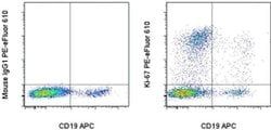
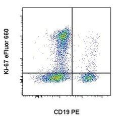
- Applications Reported: This SolA15 antibody has been reported for use in intracellular staining followed by flow cytometric analysis
- Applications Tested: This SolA15 antibody has been tested by intracellular staining followed by flow cytometric analysis of stimulated mouse splenocytes using the Foxp3/Transcription Factor Staining Buffer Set (Product No
- 00-5523-00) and protocol
- Please refer to Staining Intracellular Antigens for Flow Cytometry, Protocol B: One step protocol for intracellular (nuclear) proteins located at thermofisher.com
- This may be used at less than or equal to 0.5 μg per test
- Ki-67 is a nuclear protein that is expressed during various stages in the cell cycle, particularly during late G1, S, G2, and M phases
- The protein has a forkhead associated domain (FHA) through which it associates with euchromatin at the perichromosomal layer, the centromeric heterochromatin, and the nucleolus
- Ki-67 is shown to have a cell cycle dependent topographical distribution with perinucleolar expression at G1, expression in the nuclear matrix at G2, and expression on the chromosomes during M phase
- Ki-67 is commonly used as a proliferation marker because it is not detected in G0 cells, but increases steadily from G1 through mitosis
- Ki-67 antibodies are useful in establishing the cell growing fraction in neoplasms
- In neoplastic tissues, the prognostic value is comparable to the tritiated thymidine-labelling index
- The correlation between low Ki-67 index and histologically low-grade tumors is strong
- Ki-67 is routinely used as a neuronal marker of cell cycling and proliferation.