p53 Antibody (TRP/817), Novus Biologicals™
Mouse Monoclonal Antibody
Manufacturer: Fischer Scientific
The price for this product is unavailable. Please request a quote
Antigen
p53
Concentration
0.2mg/mL
Applications
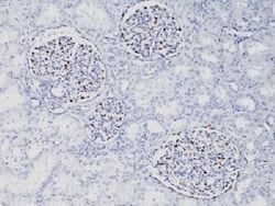
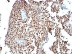
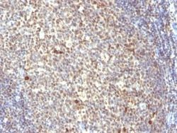
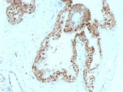
Western Blot, Flow Cytometry, Immunohistochemistry (Paraffin), Immunofluorescence, KnockDown
Conjugate
Unconjugated
Host Species
Mouse
Research Discipline
Apoptosis, Cancer, Cell Cycle and Replication, Cellular Markers, Checkpoint signaling, Core ESC Like Genes, DNA Double Strand Break Repair, DNA Repair, HIF Target Genes, Hypoxia, Neuroscience, Neurotransmission, p53 Pathway, Stem Cell Markers, Transcription Factors and Regulators, Tumor Suppressors
Formulation
10mM PBS and 0.05% BSA with 0.05% Sodium Azide
Gene ID (Entrez)
7157
Immunogen
Recombinant human TP53 protein
Primary or Secondary
Primary
Content And Storage
Store at 4C.
Molecular Weight of Antigen
53 kDa
Clone
TRP/817
Dilution
Western Blot 0.5 - 1.0 ug/ml, Flow Cytometry 0.5 - 1 ug/million cells in 0.1 ml, Immunohistochemistry-Paraffin 0.25 - 0.5 ug/ml, Immunofluorescence 0.5 - 1.0 ug/ml, Knockout Validated 0.5 ug/ml
Classification
Monoclonal
Form
Purified
Regulatory Status
RUO
Target Species
Human
Gene Alias
Antigen NY-CO-13, FLJ92943, LFS1TRP53, p53, p53 tumor suppressor, P53cellular tumor antigen p53, Phosphoprotein p53, transformation-related protein 53, tumor protein p53, Tumor suppressor p53
Gene Symbols
TP53
Isotype
IgG2b κ
Purification Method
Protein A or G purified
Test Specificity
Recognizes a 53kDa protein, which is identified as p53 suppressor gene product. It reacts with the mutant as well as the wild form of p53 protein. p53 is a tumor suppressor gene expressed in a wide variety of tissue types and is involved in regulating cell growth, replication, and apoptosis. It binds to MDM2, SV40 T antigen and human papilloma virus E6 protein. Positive nuclear staining with p53 antibody has been reported to be a negative prognostic factor in breast carcinoma, lung carcinoma, colorectal, and urothelial carcinoma. Anti-p53 positivity has also been used to differentiate uterine serous carcinoma from endometrioid carcinoma as well as to detect intratubular germ cell neoplasia. Mutations involving p53 are found in a wide variety of malignant tumors, including breast, ovarian, bladder, colon, lung, and melanoma.
Related Products


Description
- Ensure accurate, reproducible results in Western Blot, Flow Cytometry, Immunohistochemistry (Paraffin), Immunofluorescence p53 Monoclonal specifically detects p53 in Human samples
- It is validated for Western Blot, Immunohistochemistry, Immunohistochemistry-Paraffin, Knockout Validated, Knockdown Validated.