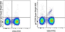
TCR alpha/beta Monoclonal Antibody (IP26), Super Bright™ 600, eBioscience™, Invitrogen™
Manufacturer: eBioscience
Select a Size
| Pack Size | SKU | Availability | Price |
|---|---|---|---|
| Each of 1 | 63-998-642-Each-of-1 | In Stock | ₹ 34,176.00 |
63-998-642 - Each of 1
In Stock
Quantity
1
Base Price: ₹ 34,176.00
GST (18%): ₹ 6,151.68
Total Price: ₹ 40,327.68
Antigen
TCR alpha/beta
Classification
Monoclonal
Concentration
5 μL/Test
Formulation
PBS with BSA and 0.09% sodium azide; pH 7.2
Gene Accession No.
P01848, P04435, P0DSE1
Gene Symbols
TRA, TRAC, TRB, TRBV7-9
Purification Method
Affinity chromatography
Regulatory Status
RUO
Gene ID (Entrez)
28589, 28755, 6955, 6957
Content And Storage
4° C, store in dark, DO NOT FREEZE!
Form
Liquid
Applications
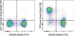
Flow Cytometry
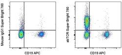
Clone
IP26
Conjugate
Super Bright 600
Gene
TRAC
Gene Alias
FLJ22602; IMD7; LOC290071; MGC117436; MGC22624; MGC23964; MGC71411; RATTCB; RATTCBC1; RGD1359684; similar to RIKEN cDNA A430107P09 gene; T cell receptor beta locus; T3/TCR complex; TCB; TCBC1; t-cell antigen receptor; T-cell receptor alpha constant; T-cell receptor beta chain; T-cell receptor V alpha; tcr alpha; TCR alpha/ beta; TCR beta; TCRA; Tcrb; TRA; Tra29; TRAC; TRB; TRB@; TRCA
Host Species
Mouse
Quantity
100 Tests
Primary or Secondary
Primary
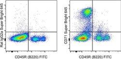
Target Species
Human
Product Type
Antibody
Isotype
IgG1 κ
Related Products


Description
- Description: The HIR2 monoclonal antibody reacts with human glycophorin A, sialoglycoproteins expressed by erythroid precursors and mature circulating red cells
- Applications Reported: This HIR2 antibody has been reported for use in flow cytometric analysis
- Applications Tested: This HIR2 antibody has been pre-diluted and tested by flow cytometric analysis of normal human peripheral blood cells
- Binding of this antibody to red cells at high antibody concentration causes cell agglutination
- This may be used at 5 μL (0.015 μg) per test
- A test is defined as the amount (μg) of antibody that will stain a cell sample in a final volume of 100 μL
- Cell number should be determined empirically but can range from 10^5 to 10^8 cells/test
- Super Bright 600 is a tandem dye that can be excited with the violet laser line (405 nm) and emits at 600 nm
- We recommend using a 610/20 bandpass filter
- Please make sure that your instrument is capable of detecting this fluorochrome
- When using two or more Super Bright dye-conjugated antibodies in a staining panel, it is recommended to use Super Bright Staining Buffer (Product No
- SB-4400) to minimize any non-specific polymer interactions
- Please refer to the datasheet for Super Bright Staining Buffer for more information
- Light sensitivity: This tandem dye is sensitive to photo-induced oxidation
- Please protect this vial and stained samples from light
- Fixation: Samples can be stored in IC Fixation Buffer (Product No
- The T cell antigen receptor (TCR) consists of a ligand-specific alpha/beta heterodimer non-covalently associated with five invariant chains including the CD3 gamma/delta/eta and zeta subunits, all of which are required for efficient surface expression
- T cell activation through the TCR induces cellular differentiation and/or proliferation and the production of lymphokines and cytokines
- Both the CD3 and TCR zeta subunits are proposed to be responsible for the intracellular signal transduction events
- Majority of T cells present in the blood, lymph and secondary lymphoid organs express TCR alpha/beta heterodimers, whereas the T cells expressing TCR gamma/delta heterodimers are localized mainly in epithelial tissues and at the sites of infection
- The subunits of TCR heterodimers are covalently bonded and associate with the CD3 subunits in the endoplasmic reticulum to form functional TCR-CD3 complex
- Lack of expression of any of the chains is sufficient to stop cell surface expression
- The ability of T cell receptors (TCR) to discriminate foreign from self-peptides presented by major histocompatibility complex (MHC) class II molecules is essential for an effective adaptive immune response
- TCR recognition of self-peptides has been linked to autoimmune disease
- Mutant self-peptides have been associated with tumors
- Engagement of TCRs by a family of bacterial toxins know as superantigens has been responsible for toxic shock syndrome
- Autoantibodies to V beta segments of T cell receptors have been isolated from patients with rheumatoid arthritis (RA) and systemic lupus erythematosus (SLE)
- The autoantibodies block TH1-mediated inflammatory auto-destructive reactions and are believed to be a method by which the immune system compensates for disease (ref5)
- T Cell and TCR Diversity Most human T cells express the TCR alpha-beta and either CD4 or CD8 molecule (single positive, SP)
- A small number of T cells lack both CD4 and CD8 (double negative, DN)
- Increased percentages of alpha-beta DN T cells have been identified in some autoimmune and immunodeficiency disorders
- Gamma-delta T cells are primarily found within the epithelium
- They show less TCR diversity and recognize antigens differently than alpha-beta T cells
- Subsets of gamma-delta T cells have shown antitumor and immunoregulatory activity.