7068538
CD279 (PD-1) Monoclonal Antibody (RMP1-30), FITC, eBioscience™, Invitrogen™
Rat Monoclonal Antibody
Manufacturer: Fischer Scientific
The price for this product is unavailable. Please request a quote
Antigen
CD279 (PD-1)
Concentration
0.5 mg/mL
Classification
Monoclonal
Form
Liquid
Regulatory Status
RUO
Formulation
PBS with 0.09% sodium azide; pH 7.2
Gene Alias
CD279; EGK_05005; hPD1; hPD-1; hPD-l; hSLE1; Ly101; mPD-1; PD1; PD-1; Pdc1; Pdcd1; programmed cell death 1; programmed cell death 1 protein; programmed cell death protein 1; programmed cell death protein 1-like; programmed death 1; Protein PD1; protein PD-1; sCD279; SLEB2; soluble CD279; systemic lupus erythematosus susceptibility 2
Gene Symbols
Pdcd1
Primary or Secondary
Primary
Content And Storage
4° C, store in dark, DO NOT FREEZE!
Gene
Pdcd1
Clone
RMP1-30
Applications
Flow Cytometry
Conjugate
FITC
Host Species
Rat
Target Species
Mouse
Gene Accession No.
Q02242
Gene ID (Entrez)
18566
Isotype
IgG2b κ
Purification Method
Affinity chromatography
Product Type
Antibody
Related Products


Description
- Description: The RMP1-30 antibody reacts with mouse PD-1 (programmed death-1), a 55 kDa member of the Ig superfamily
- PD-1 contains the immunoreceptor tyrosine-based inhibitory motif (ITIM) and plays a key role in peripheral tolerance and autoimmune disease in mice
- PD-1 is expressed mainly on activated T and B lymphocytes
- Two novel B7 Family members have been identified as PD-1 ligands, PD-L1 (B7-H1) and PD-L2 (B7-DC)
- Evidence reported to date suggests overlapping functions for these ligands and their constitutive expression on some normal tissues and upregulation on activated antigen-presenting cells
- RMP1-30 does not block the binding of either B7-H1-Ig or B7-DC-Ig to PD-1 transfectants
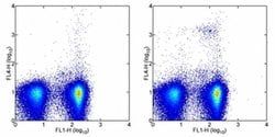
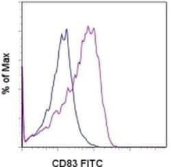
- Applications Reported: The RMP1-30 antibody has been reported for use in flow cytometric analysis
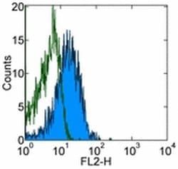
- Applications Tested: The RMP1-30 antibody has been tested by flow cytometric analysis of Con A-stimulated mouse splenocytes
- This can be used at less than or equal to 0.5 μg per test
- A test is defined as the amount (μg) of antibody that will stain a cell sample in a final volume of 100 μL
- Cell number should be determined empirically but can range from 10^5 to 10^8 cells/test
- It is recommended that the antibody be carefully titrated for optimal performance in the assay of interest
- Excitation: 488 nm; Emission: 520 nm; Laser: Blue Laser
- Filtration: 0.2 μm post-manufacturing filtered
- Cell-mediated immune responses are initiated by T lymphocytes that are themselves stimulated by cognate peptides bound to MHC molecules on antig en-presenting cells (APC)
- T-cell activation is generally self-limited as activated T cells express receptors such as PD-1 (also known as PDCD-1) that mediate inhibitory signals from the APC
- PD-1 can bind two different but related ligands, PDL-1 and PDL-2
- Upon binding to either of these ligands, signals generated by PD-1 inhibit the activation of the immune response in the absence of "edanger signals"e such as LPS or other molecules associated with bacteria or other pathogens
- Evidence for this is seen in PD1-null mice who exhibit hyperactivated immune systems and autoimmune diseases
- Despite its predicted molecular weight, PD-1 often migrates at higher molecular weight in SDS-PAGE.