7068532
CD279 (PD-1) Monoclonal Antibody (MIH4), PE, eBioscience™, Invitrogen™
Mouse Monoclonal Antibody
Manufacturer: Fischer Scientific
The price for this product is unavailable. Please request a quote
Antigen
CD279 (PD-1)
Concentration
5 μL/Test
Classification
Monoclonal
Form
Liquid
Regulatory Status
RUO
Formulation
PBS with 0.2% BSA and 0.09% sodium azide; pH 7.2
Gene Alias
CD279; EGK_05005; hPD1; hPD-1; hPD-l; hSLE1; Ly101; mPD-1; PD1; PD-1; Pdc1; Pdcd1; programmed cell death 1; programmed cell death 1 protein; programmed cell death protein 1; programmed cell death protein 1-like; programmed death 1; Protein PD1; protein PD-1; sCD279; SLEB2; soluble CD279; systemic lupus erythematosus susceptibility 2
Gene Symbols
Pdcd1
Primary or Secondary
Primary
Content And Storage
4° C, store in dark, DO NOT FREEZE!
Gene
Pdcd1
Clone
MIH4
Applications
Flow Cytometry
Conjugate
PE
Host Species
Mouse
Target Species
Human
Gene Accession No.
Q15116
Gene ID (Entrez)
5133
Isotype
IgG1 κ
Purification Method
Affinity chromatography
Product Type
Antibody
Related Products


Description
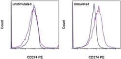
- Description: The MIH4 monoclonal antibody reacts with the human PD-1 (programmed death-1), a 55 kDa member of the immunoglobulin superfamily
- PD-1 contains the immunoreceptor tyrosine-based inhibitory motif (ITIM) and plays a key role in peripheral tolerance and autoimmune disease
- PD-1 is expressed predominantly on activated T and B lymphocytes
- Two novel members of the B7 family have been identified as the PD-1 ligands, PD-L1 (B7-H1) and PD-L2 (B7-DC)
- Evidence reported to date suggests overlapping functions for these two PD-1 ligands and their constitutive expression on some normal tissues and upregulation on activated antigen-presenting cells
- The MIH4 antibody recognizes a different epitope than antibody clones J105
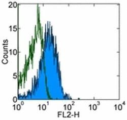
- Applications Reported: This MIH4 antibody has been reported for use in flow cytometric analysis
- Applications Tested: This MIH4 antibody has been pre-titrated and tested by flow cytometric analysis of PHA-stimulated normal human peripheral blood cells
- This can be used at 5 μL (0.5 μg) per test
- A test is defined as the amount (μg) of antibody that will stain a cell sample in a final volume of 100 μL
- Cell number should be determined empirically but can range from 10^5 to 10^8 cells/test
- Excitation: 488-561 nm; Emission: 578 nm; Laser: Blue Laser, Green Laser, Yellow-Green Laser
- Filtration: 0.2 μm post-manufacturing filtered
- Cell-mediated immune responses are initiated by T lymphocytes that are themselves stimulated by cognate peptides bound to MHC molecules on antig en-presenting cells (APC)
- T-cell activation is generally self-limited as activated T cells express receptors such as PD-1 (also known as PDCD-1) that mediate inhibitory signals from the APC
- PD-1 can bind two different but related ligands, PDL-1 and PDL-2
- Upon binding to either of these ligands, signals generated by PD-1 inhibit the activation of the immune response in the absence of "edanger signals"e such as LPS or other molecules associated with bacteria or other pathogens
- Evidence for this is seen in PD1-null mice who exhibit hyperactivated immune systems and autoimmune diseases
- Despite its predicted molecular weight, PD-1 often migrates at higher molecular weight in SDS-PAGE.