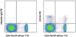
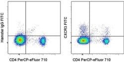
CD183 (CXCR3) Monoclonal Antibody (CEW33D), PE, eBioscience™, Invitrogen™
Mouse Monoclonal Antibody
Manufacturer: Fischer Scientific
The price for this product is unavailable. Please request a quote
Antigen
CD183 (CXCR3)
Concentration
5 μL/Test
Classification
Monoclonal
Form
Liquid
Regulatory Status
RUO
Formulation
PBS with 0.2% BSA and 0.09% sodium azide; pH 7.2
Gene Alias
an; C Cmotif chemokine; C X C motif chemokine; CC motif chemokine; CCmotif chemokine; CD182; CD183; chemokine (C-X-C motif) receptor 3; chemokine receptor 3; CKR-L2; Cmkar3; CXC; C-X-C chemokine receptor type 3; CXC motif chemokine; C-X-C motif chemokine receptor 3; Cxcr3; CXC-R3; CXCR-3; G protein-coupled receptor 9; GPR9; Interferon-inducible protein 10 receptor; IP10; IP10 receptor; IP-10 receptor; IP10-R; Mig receptor; MigR; Mig-R
Gene Symbols
CXCR3
Primary or Secondary
Primary
Content And Storage
4° C, store in dark, DO NOT FREEZE!
Gene
CXCR3
Clone
CEW33D
Applications
Flow Cytometry
Conjugate
PE
Host Species
Mouse
Target Species
Human
Gene Accession No.
P49682
Gene ID (Entrez)
2833
Isotype
IgG1 κ
Purification Method
Affinity chromatography
Product Type
Antibody
Related Products


Description
- Description: The CEW33D monoclonal antibody reacts with human CD183
- CD183, also known as CXCR3, is a G protein-coupled chemokine receptor that interacts with ligands CXCL9 (MIG), CXCL10 (IP-10), and CXCL11 (I-TAC)
- Strongly associated with type 1 immunity, CD183 is induced in naive T cells upon activation and remains upregulated in T helper type (Th)1 cells, CD8 effector cells, NK cells and NKT cells
- CD183-ligand interactions mediate infiltration of inflamed tissues in normal type 1 immune responses as well as in many inflammatory and autoimmune diseases
- CD183 is also expressed on some B cells and plasmacytoid DC
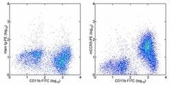
- Applications Reported: This CEW33D antibody has been reported for use in flow cytometric analysis
- Applications Tested: This CEW33D antibody has been pre-titrated and tested by flow cytometric analysis of normal human peripheral blood cells
- This can be used at 5 μL (0.25 μg) per test
- A test is defined as the amount (μg) of antibody that will stain a cell sample in a final volume of 100 μL
- Cell number should be determined empirically but can range from 10^5 to 10^8 cells/test
- Excitation: 488-561 nm; Emission: 578 nm; Laser: Blue Laser, Green Laser, Yellow-Green Laser
- Filtration: 0.2 μm post-manufacturing filtered
- This gene encodes a G protein-coupled receptor with selectivity for three chemokines, termed IP10 (interferon-g-inducible 10 kDa protein), Mig (monokine induced by interferon-g) and I-TAC (interferon-inducible T cell a-chemoattractant)
- IP10, Mig and I-TAC belong to the structural subfamily of CXC chemokines, in which a single amino acid residue separates the first two of four highly conserved Cys residues
- Binding of chemokines to this protein induces cellular responses that are involved in leukocyte traffic, most notably integrin activation, cytoskeletal changes and chemotactic migration
- Inhibition by Bordetella pertussis toxin suggests that heterotrimeric G protein of the Gi-subclass couple to this protein
- Signal transduction has not been further analyzed but may include the same enzymes that were identified in the signaling cascade induced by other chemokine receptors
- As a consequence of chemokine-induced cellular desensitization (phosphorylation-dependent receptor internalization), cellular responses are typically rapid and short in duration
- Cellular responsiveness is restored after dephosphorylation of intracellular receptors and subsequent recycling to the cell surface
- This gene is prominently expressed in in vitro cultured effector/memory T cells, and in T cells present in many types of inflamed tissues
- In addition, IP10, Mig and I-TAC are commonly produced by local cells in inflammatory lesion, suggesting that this gene and its chemokines participate in the recruitment of inflammatory cells
- Therefore, this protein is a target for the development of small molecular weight antagonists, which may be used in the treatment of diverse inflammatory diseases
- Multiple transcript variants encoding different isoforms have been found for this gene.