7139417
Arginase 1 Monoclonal Antibody (A1exF5), PE-Cyanine7, eBioscience™, Invitrogen™
Rat Monoclonal Antibody
Manufacturer: Fischer Scientific
The price for this product is unavailable. Please request a quote
Antigen
Arginase 1
Concentration
0.2 mg/mL
Classification
Monoclonal
Form
Liquid
Regulatory Status
RUO
Formulation
PBS with 0.09% sodium azide; pH 7.2
Gene Alias
AI; A-I; AI type I arginase; AI256583; Arg1; Arg-1; Arginase; arginase 1; arginase 1 liver; arginase 1, liver; arginase I; arginase, liver; Arginase1; arginase-1; HGNC:663; Liver Arginase; Liver-type arginase; PGIF; similar to arginase, type I; Type 1 Arginase; type I arginase
Gene Symbols
ARG1
Isotype
IgG2a κ
Purification Method
Affinity chromatography
Product Type
Antibody
Clone
A1exF5
Applications
Flow Cytometry
Conjugate
PE-Cyanine7
Host Species
Rat
Target Species
Human, Mouse
Gene Accession No.
P05089, Q61176
Gene ID (Entrez)
11846, 383
Immunogen
E.coli-derived recombinant mouse Arginase 1
Primary or Secondary
Primary
Content And Storage
4° C, store in dark, DO NOT FREEZE!
Gene
ARG1
Related Products
Description
- Description: The monoclonal antibody A1exF5 recognizes both human and mouse Arginase 1, a cytosolic enzyme (Arg1)
- This A1exF5 clone is compatible with both, the standard intracellular protocols, and the Foxp3/Transcription Factor Staining Buffer Set
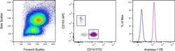
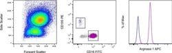
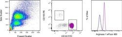
- Applications Reported: This A1exF5 antibody has been reported for use in flow cytometric analysis
- Applications Tested: This A1exF5 antibody has been tested by flow cytometric analysis of normal human peripheral blood cells using the Intracellular Fixation & Permeabilization Buffer Set (Product No
- 88-8824) and protocol
- Please refer to Best Protocols: Protocol A: Two step protocol for (cytoplasmic) intracellular proteins located under the Resources Tab online
- This may be used at less than or equal to 0.5 μg per test
- A test is defined as the amount (μg) of antibody that will stain a cell sample in a final volume of 100 μL
- Cell number should be determined empirically but can range from 10^5 to 10^8 cells/test
- It is recommended that the antibody be carefully titrated for optimal performance in the assay of interest
- Light sensitivity: This tandem dye is sensitive to photo-induced oxidation
- Please protect this vial and stained samples from light
- Fixation: Samples can be stored in IC Fixation Buffer (Product No
- 00-8222) (100 μL of cell sample + 100 μL of IC Fixation Buffer) or 1-step Fix/Lyse Solution (Product No
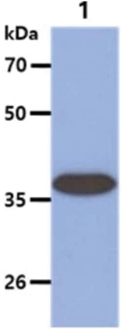
- Arginase-1 (Arg1) is a 35 kDa enzyme converting L-arginine to urea and L-ornithine, which is the final step in the urea cycle
- The resulting polyamines are important for cell proliferation and removal of toxins that arise from protein degradation
- By degrading arginine, Arginase 1 deprives NO synthase of its substrate and down-regulates nitric oxide production
- In both human and mouse, Arginase 1 is expressed in the liver, neutrophils, myeloid derived suppressor cells (MDSC) and neural stem cells
- In human, expression in blood neutrophils but not in CCR3+ granulocytes has been reported
- In mice, expression of Arginase 1 is one of the hallmarks of alternatively activated macrophages (M2a)
- Arginase-1 may be expressed in the myeloid cells infiltrating tumors, and is typically found in the majority of hepatocellular carcinomas
- Defects in Arginase 1 are the cause of argininemia, an autosomal recessive disorder characterized by hyperammonemia.