CD63 (LAMP3), Mouse anti-Human, Clone: ME491, Millipore Sigma™
Mouse Monoclonal Antibody
Manufacturer: Fischer Scientific
The price for this product is unavailable. Please request a quote
Antigen
CD63 (LAMP3)
Dilution
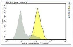
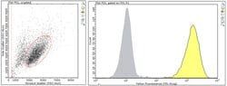
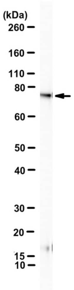
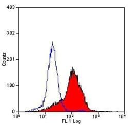
Immunohistochemistry (Paraffin) Analysis: A 1:250 dilution from a representative lot detected CD63 (LAMP3) in human spleen and human bone marrow tissue sections.Immunocytochemistry Analysis: A representative lot detected CD63 (LAMP3) in Immunocytochemistry applications (Atkinson, B., et. al. (1984). Cancer Res. 44(6):2577-81).Flow Cytometry Analysis: A representative lot detected CD63 (LAMP3) in Flow Cytometry applications (Li, J., et. al. (2003). J Immunol. 171(6):2922-9).Western Blotting Analysis: A representative lot detected CD63 (LAMP3) in Western Blotting applications (Smith, M., et. al. (1997). Melanoma Res. 7 Suppl 2:S163-70).Immunohistochemistry Analysis: A representative lot detected CD63 (LAMP3) in Immunohistochemistry applications (Li, J., et. al. (2003). J Immunol. 171(6):2922-9).
Classification
Monoclonal
Form
Purified
Regulatory Status
RUO
Target Species
Human
Gene Alias
CD63 antigen;Granulophysin;Lysosomal-associated membrane protein 3;LAMP-3;Melanoma-associated antigen ME491;OMA81H;Ocular melanoma-associated antigen;Tetraspanin-30;Tspan-30
Gene Symbols
CD63;MLA1;TSPAN30
Isotype
IgG1 κ
Purification Method
Protein G purified
Test Specificity
Clone ME491 specifically detects CD63 (LAMP-3) in human cells.
Clone
ME491
Applications
Flow Cytometry, Immunocytochemistry, Immunohistochemistry (Paraffin), Western Blot
Conjugate
Unconjugated
Host Species
Mouse
Research Discipline
Inflammation & Immunology
Formulation
Purified mouse monoclonal antibody IgG1 in buffer containing 0.1 M Tris-Glycine (pH 7.4), 150 mM NaCl with 0.05% sodium azide.
Gene ID (Entrez)
NP_001244318
Immunogen
Clear supernatant from SK-Mel-23 cell lysate.
Primary or Secondary
Primary
Content And Storage
Stable for 1 year at 2-8°C from date of receipt.
Related Products


Description
- Anti-CD63 (LAMP3), clone ME491, Cat
- No
- MABF2159, is a mouse monoclonal antibody that detects CD63 antigen and has been tested for use in Flow Cytometry, Immunocytochemistry, Immunohistochemistry (Paraffin), and Western Blotting
- CD63 antigen (UniProt: P08962; also known as Granulophysin, Lysosomal-associated membrane protein 3, LAMP-3, Melanoma-associated antigen ME491, OMA81H, Ocular melanoma-associated antigen, Tetraspanin-30, Tspan-30, CD63) is encoded by the CD63 (also known as MLA1, TSPAN30) gene (Gene ID: 967) in human
- CD63 is a multi-pass membrane protein of the tetraspan family that is found on endosome, lysosome, and plasma membranes
- CD63 has been detected in platelets, Dysplastic nevi benign moles), radial growth phase primary melanomas, hematopoietic cells, and in tissue macrophages
- In melanoma cells it is involved in their motility and adhesion
- CD63 also plays a role in the adhesion of leukocytes onto endothelial cells
- It is reported to play a role in the activation of ITGB1 and integrin signaling, leading to the activation of AKT, FAK/PTK2 and MAP kinases and promote cell survival, reorganization of the actin cytoskeleton, cell adhesion, spreading and migration
- CD63 is a highly N-glycosylated protein with three asparagine glycosylation sites (aa 130, 150, 172) and its ribophorin II (RPN2)-mediated glycosylation has been linked to breast cancer
- Overexpression of CD63 has been observed in esophageal cancer that is negatively correlated with tumor stage and lymph node metastasis
- Lack of expression of CD63 in platelets has been observed in a patient with Hermansky-Pudlak syndrome (HPS), an autosomal recessive disorder that is characterized by oculocutaneous albinism, bleeding due to platelet storage pool deficiency, and lysosomal storage defects
- This antibody (clone ME491) is shown to react with human primary and to some extent with metastatic melanoma tissues
- (Ref.: Lai, X., et al
- (2017)
- Oncol
- Let
- 13(6); 4245-4251; Tominaga, N., et al
- (2014)
- Mol
- Cancer 13; 134; Smith, M., et al
- (1997)
- Melanoma Res
- 7 (Suppl
- 2), 163-170).