phospho-cytokeratin-18 (K18) (Ser33), Mouse anti-Human, Clone: IB4, Millipore Sigma™
Manufacturer: Fischer Scientific
Select a Size
| Pack Size | SKU | Availability | Price |
|---|---|---|---|
| Each of 1 | MABT875100-Each-of-1 | In Stock | ₹ 42,969.20 |
MABT875100 - Each of 1
In Stock
Quantity
1
Base Price: ₹ 42,969.20
GST (18%): ₹ 7,734.456
Total Price: ₹ 50,703.656
Antigen
phospho-cytokeratin-18 (K18) (Ser33)
Classification
Monoclonal
Conjugate
Unconjugated
Formulation
Purified mouse monoclonal antibody IgG1 in buffer containing 0.1 M Tris-Glycine (pH 7.4), 150 mM NaCl with 0.05% sodium azide.
Gene Symbols
KRT18;CYK18;PIG46
Immunogen
KLH-conjugated linear peptide corresponding to 13 amino acids surrounding phosphorylated serine 33 (initiator methionine removed) from the N-terminal region of human cytokeratin-18.
Quantity
100 μg
Research Discipline
Cell Structure
Gene ID (Entrez)
NP_000215
Target Species
Human
Form
Purified
Applications
Immunocytochemistry, Immunofluorescence, Immunoprecipitation, Western Blot
Clone
IB4
Dilution
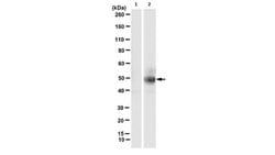
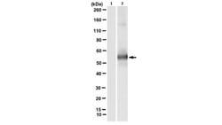
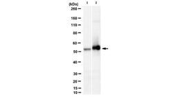
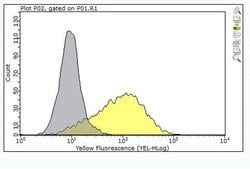
Immunocytochemistry Analysis: A representative lot detected phospho-cytokeratin-18 (K18) (Ser33) in immunocytochemistry applications (Ku, N.O., et. al. (1998). EMBO J. 17(7):1892-906).Immunoprecipitation Analysis: A representative lot immunoprecipitated phospho-cytokeratin-18 (K18) (Ser33) in immunoprecipitation applications (Ku, N.O., et. al. (1998). EMBO J. 17(7):1892-906).Immunofluorescence Analysis: A representative lot detected phospho-cytokeratin-18 (K18) (Ser33) in Immunofluorescence applications (Ku, N.O., et. al. (1998). EMBO J. 17(7):1892-906).Western Blotting Analysis: A representative lot detected phospho-cytokeratin-18 (K18) (Ser33) in Western Blotting applications (Yoon, K.H., et. al. (2001). J Cell Biol. 153(3):503-16).
Gene Alias
Keratin type I cytoskeletal 18;Cell proliferation-inducing gene 46 protein;CK-18;Keratin-18;K18
Host Species
Mouse
Purification Method
Protein G purified
Regulatory Status
RUO
Primary or Secondary
Primary
Test Specificity
Clone IB4 specifically detects human cytokeratin-18 phosphorylated on serine 33.
Content And Storage
Stable for 1 year at 2-8°C from date of receipt.
Isotype
IgG1 κ
Related Products


Description
- Anti-phospho-cytokeratin-18 (K18) (Ser33), clone IB4, Cat
- No
- MABT875, is a mouse monoclonal antibody that detects type I cytoskeletal-18 and has been tested for use in Immunocytochemistry, Immunofluorescence, Immunoprecipitation, and Western Blotting
- Keratin, type I cytoskeletal 18 (UniProt: P05783; also known as Cell proliferation-inducing gene 46 protein, Cytokeratin-18, CK-18, Keratin-18, K18) is encoded by the KRT18 (also known as CYK18, PIG46) gene (Gene ID: 3875) in human
- Cytokeratin-18 is a type I, acidic, protein that is expressed in colon, placenta, and liver
- Higher expression levels have been reported in lymph nodes of breast carcinoma
- Cytokeratin-18 is involved in the uptake of thrombin-antithrombin complexes by hepatic cells
- Upon phosphorylation, it plays a role in filament reorganization
- It is proteolytically cleaved by caspases during epithelial cell apoptosis and this cleavage is shown to occur at Asp-238
- Phosphorylation of cytokeratin-18 at serine 33 is shown to increase during mitosis in cultured cells and in regenerating liver
- Serine 33 phosphorylation is considered to be essential for its association with 14-3-3 proteins and has a role in keratin organization and distribution
- Cytokeratin-18 can undergo O-glycosylation, which results in its increased solubility and reduction in its stability
- Mutations in KRT18 gene are known to cause liver cirrhosis characterized by severe panlobular liver-cell swelling with Mallory body formation, prominent pericellular fibrosis, and marked deposits of copper.