58-044-182
CD44 Monoclonal Antibody (IM7), Alexa Fluor™ 532, eBioscience™, Invitrogen™
Manufacturer: LIFE TECHNOLOGIES
Select a Size
| Pack Size | SKU | Availability | Price |
|---|---|---|---|
| Each of 1 | 58-044-182-Each-of-1 | In Stock | ₹ 33,197.00 |
58-044-182 - Each of 1
In Stock
Quantity
1
Base Price: ₹ 33,197.00
GST (18%): ₹ 5,975.46
Total Price: ₹ 39,172.46
Antigen
CD44
Classification
Monoclonal
Concentration
0.2 mg/ml
Formulation
PBS with 0.09% sodium azide; pH 7.2
Gene Accession No.
P15379, P16070
Host Species
Rat
Quantity
100 μg
Primary or Secondary
Primary
Target Species
Human, Mouse
Product Type
Antibody
Isotype
IgG2b κ
Applications
Flow Cytometry
Clone
IM7
Conjugate
Alexa Fluor 532
Gene
CD44
Gene Symbols
Cd44
Purification Method
Affinity chromatography
Regulatory Status
RUO
Gene ID (Entrez)
12505, 960
Content And Storage
4° C, store in dark, DO NOT FREEZE!
Form
Liquid
Related Products

Description
- Description: The IM7 monoclonal antibody reacts with all isoforms of mouse CD44 (Pgp-1)
- CD44 is expressed by hematopoietic and non-hematopoietic cells
- Bone marrow myeloid cells and memory T cells highly express this antigen and peripheral B and T cells can upregulate the expression of CD44
- CD44 functions as an adhesion molecule through its binding to hyaluronate, an extracellular matrix component
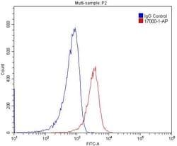
- Applications Reported: This IM7 antibody has been reported for use in flow cytometric analysis
- Applications Tested: This IM7 antibody has been tested by flow cytometric analysis of mouse splenocytes
- This may be used at less than or equal to 0.5 μg per test
- A test is defined as the amount (μg) of antibody that will stain a cell sample in a final volume of 100 μL
- Cell number should be determined empirically but can range from 10^5 to 10^8 cells/test
- It is recommended that the antibody be carefully titrated for optimal performance in the assay of interest
- Alexa Fluor 532 is excited with the Green laser (532 nm) and emits at 561 nm
- This cannot be used with the Yellow-Green laser (561 nm)
- We recommend using a 560/14 band pass filter
- Please make sure that your instrument is capable of detecting this fluorochrome
- Excitation: 532 nm; Emission: 561 nm; Laser: Green Laser CD44 cell surface antigen is a 100 kDa type 1 transmembrane glycoprotein widely expressed on human leucocytes, white matter of the brain and by some epithelial cells of the intestine and breast
- Several isoforms of CD44 exist, including the predomit CD44H isoform detected in many normal tissues
- CD44 is a receptor for hyaluronic acid (HA) and is involved in cell-cell interactions, cell adhesion and migration
- CD44 also participates in a wide variety of cellular functions including lymphocyte activation, recirculation and homing
- CD44 expression may be up-regulated upon some carcinomas, and it has been speculated that this may be related to metastatic potential
- CD44 is expressed by hematopoietic, non-hematopoietic cells, epithelial tissues, and to filopodia in cultured keratinocytes
- Further, bone marrow myeloid cells and memory T cells express CD44 at high levels, and peripheral B and T cells can upregulate the expression of CD44 in response to certain stimulatory events
- Transcripts for the CD44 gene undergo complex alternative splicing that results in many functionally distinct isoforms, however, the full-length nature of some of these variants have not been determined
- Alternative splicing is the basis for the structural and functional diversity of the CD44 protein
- Diseases associated with CD44 dysfunction include superficial keratitis and lichen sclerosus
- CD44 also may be related to tumor metastasis formation.