7143936
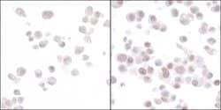
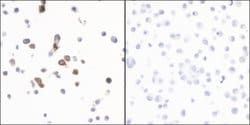
Ki-67 Recombinant Rabbit Monoclonal Antibody (BLR021E), Bethyl Laboratories
Rabbit Recombit Monoclonal Antibody
Manufacturer: Fischer Scientific
The price for this product is unavailable. Please request a quote
Antigen
Ki-67
Applications
Immunocytochemistry, Immunohistochemistry, Western Blot
Conjugate
Unconjugated
Host Species
Rabbit
Target Species
Human
Gene Accession No.
P46013
Gene ID (Entrez)
4288
Immunogen
Between 1200 and 1250
Primary or Secondary
Primary
Content And Storage
4° C
Gene
MKI67
Clone
BLR021E
Classification
Recombinant Monoclonal
Form
Liquid
Regulatory Status
RUO
Formulation
BBS with 0.1% BSA and 0.09% sodium azide; pH 8.2
Gene Alias
antigen identified by monoclonal antibody Ki-67, KIA, MIB-, MIB-1, PPP1R105, proliferation-related Ki-67 antigen, protein phosphatase 1, regulatory subunit 105
Gene Symbols
MKI67
Isotype
IgG
Purification Method
Ion-exchange chromatography
Product Type
Antibody
Related Products
Description
- The recommended shelf life for this product is 1 year from date of receipt
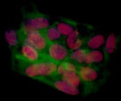
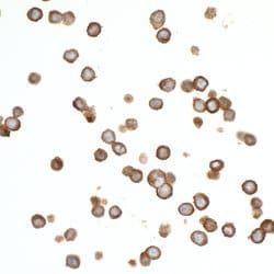
- Application Note: For ICC, epitope retrieval with citrate buffer pH 6.0 is recommended for FFPE cell sections
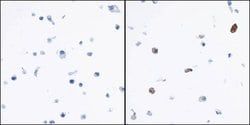
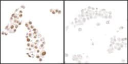
- For IHC, epitope retrieval with citrate buffer pH 6.0 is recommende Ki-67 is a nuclear protein that is expressed during various stages in the cell cycle, particularly during late G1, S, G2, and M phases
- The protein has a forkhead associated domain (FHA) through which it associates with euchromatin at the perichromosomal layer, the centromeric heterochromatin, and the nucleolus
- Ki-67 is shown to have a cell cycle dependent topographical distribution with perinucleolar expression at G1, expression in the nuclear matrix at G2, and expression on the chromosomes during M phase
- Ki-67 is commonly used as a proliferation marker because it is not detected in G0 cells, but increases steadily from G1 through mitosis
- Ki-67 antibodies are useful in establishing the cell growing fraction in neoplasms
- In neoplastic tissues, the prognostic value is comparable to the tritiated thymidine-labelling index
- The correlation between low Ki-67 index and histologically low-grade tumors is strong
- Ki-67 is routinely used as a neuronal marker of cell cycling and proliferation.