50-112-2662
CD207 (Langerin) Monoclonal Antibody (eBioL31), eBioscience™, Invitrogen™
Manufacturer: Invitrogen
Select a Size
| Pack Size | SKU | Availability | Price |
|---|---|---|---|
| Each of 1 | 50-112-2662-Each-of-1 | In Stock | ₹ 28,213.00 |
50-112-2662 - Each of 1
In Stock
Quantity
1
Base Price: ₹ 28,213.00
GST (18%): ₹ 5,078.34
Total Price: ₹ 33,291.34
Antigen
CD207 (Langerin)
Classification
Monoclonal
Concentration
0.5 mg/mL
Formulation
PBS with 0.09% sodium azide; pH 7.2
Gene Accession No.
Q8VBX4
Gene Symbols
CD207
Purification Method
Affinity chromatography
Regulatory Status
RUO
Gene ID (Entrez)
246278
Content And Storage
4° C
Form
Liquid
Applications
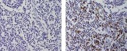
Flow Cytometry, Immunohistochemistry (Frozen), Immunoprecipitation, Western Blot
Clone
eBioL31
Conjugate
Unconjugated
Gene
CD207
Gene Alias
CD 207 antigen; CD207; CD207 antigen; CD207 antigen, langerin; CD207 molecule; CD207 molecule, langerin; CLEC4K; C-type lectin domain family 4 member K; C-type lectin domain family 4, member K; Langerhans cell specific c-type lectin; langerin; RGD1565913
Host Species
Rat
Quantity
100 μg
Primary or Secondary
Primary
Target Species
Mouse
Product Type
Antibody
Isotype
IgG2a
Related Products


Description
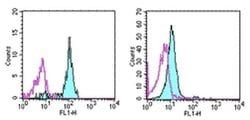
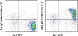
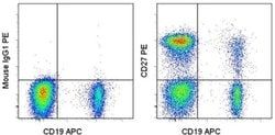
- Description: The eBioL31 monoclonal antibody reacts with mouse CD207, also known as Langerin, which is expressed in a distinct subset of dendritic cells called Langerhans cells (LC)
- Mouse CD207 is a 48 kDa C-type lectin transmembrane protein that likely plays a role in antigen recognition and uptake
- LC are located in the epidermis and upon activation, reduce CD207 expression and begin migrating through the dermis towards lymphatic vessels
- Expression of mouse CD207 in LC has been correlated to the presence of Birbeck Granules
- Furthermore, the formation of Birbeck Granules has been observed upon transfection of CD207 cDNA into fibroblast cell lines
- Western Blotting with eBioL31 reveals a 48kDa band in lysate from mouse ear epidermis, as well as in lysates from cells transfected with mouse CD207 cDNA
- Preliminary data from mouse CD207 cDNA transfected cells suggests that eBioL31 can be used for intracellular flow cytometric analysis in combination with the Foxp3/Transcription Factor Staining Bufer Set (cat
- 00-5523)
- The epitope recognized by eBioL31 is in the extracellular domain within the CRD (carbohydrate recognition domain) and therefore can be used to detect both surface and intracellular langerin
- Differences in expression has been observed between mouse strains
- Langerin (CD207) is a type II membrane-associated C-type lectin known to be expressed exclusively by Langerhans cells
- Lamgerin recognizes mannose residues via its single carbohydrate recognition domain (CRD)
- Langerin is localized not only on the cell surface, but also intracellularly in close association with Birbeck granules
- Transfection of Langerin cDNA into fibroblasts creates a compact network of membrane structures with typical features of Birbeck granules (BG)
- Langerin is thus a potent inducer of membrane superimposition and zippering leading to BG formation.